Interference Reflection Microscopy using the Olympus FluoView FV1200
Interference Reflection Microscopy (IRM) is normally used to study cellular contact sites on a glass surface without using fluorescent labels. Confocal microscope lasers are polarised, which makes them ideal for IRM. You can acquire fluorescence images at the same time as reflection images but will have to match the dichroics and emission filters to the channels you want to image.
Setting up reflection imaging on the FV1200
Click on the "light paths and dyes"  button to open the tools for setting up the light paths.
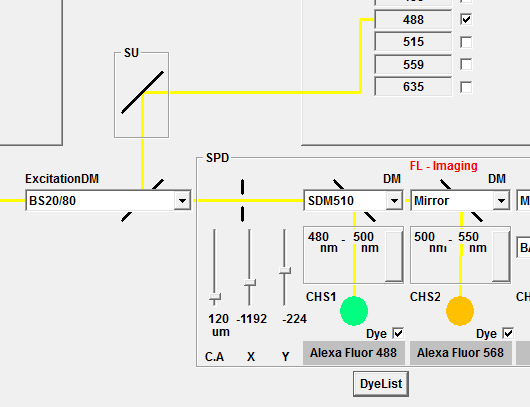
Set the ExcitationDM to BS20/80 to allow laser reflection through to the detectors. In the example below, the laser in use is 488 for both reflection and fluorescence. SDM510 directs all wavelenghts below 510nm to detector CHS1, and the emission bandwidth is set to 480-500 to allow reflection from the 488 laser through. The dichroic mirror before detector CHS2 is set up to reflect all of the remaining light to the detector, and the emission bandwith set to 500-550 to detect green fluorescence. Other lasers can be used, but you may be limited by the dichroics available.
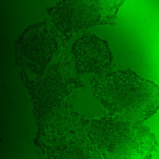
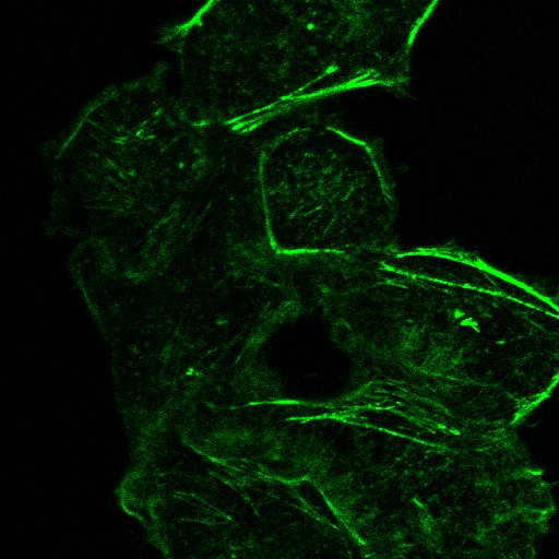
You should end up with a reflection and a confocal fluorescence image like the ones below.