Olympus FluoView FV1200
GENERAL INFORMATION
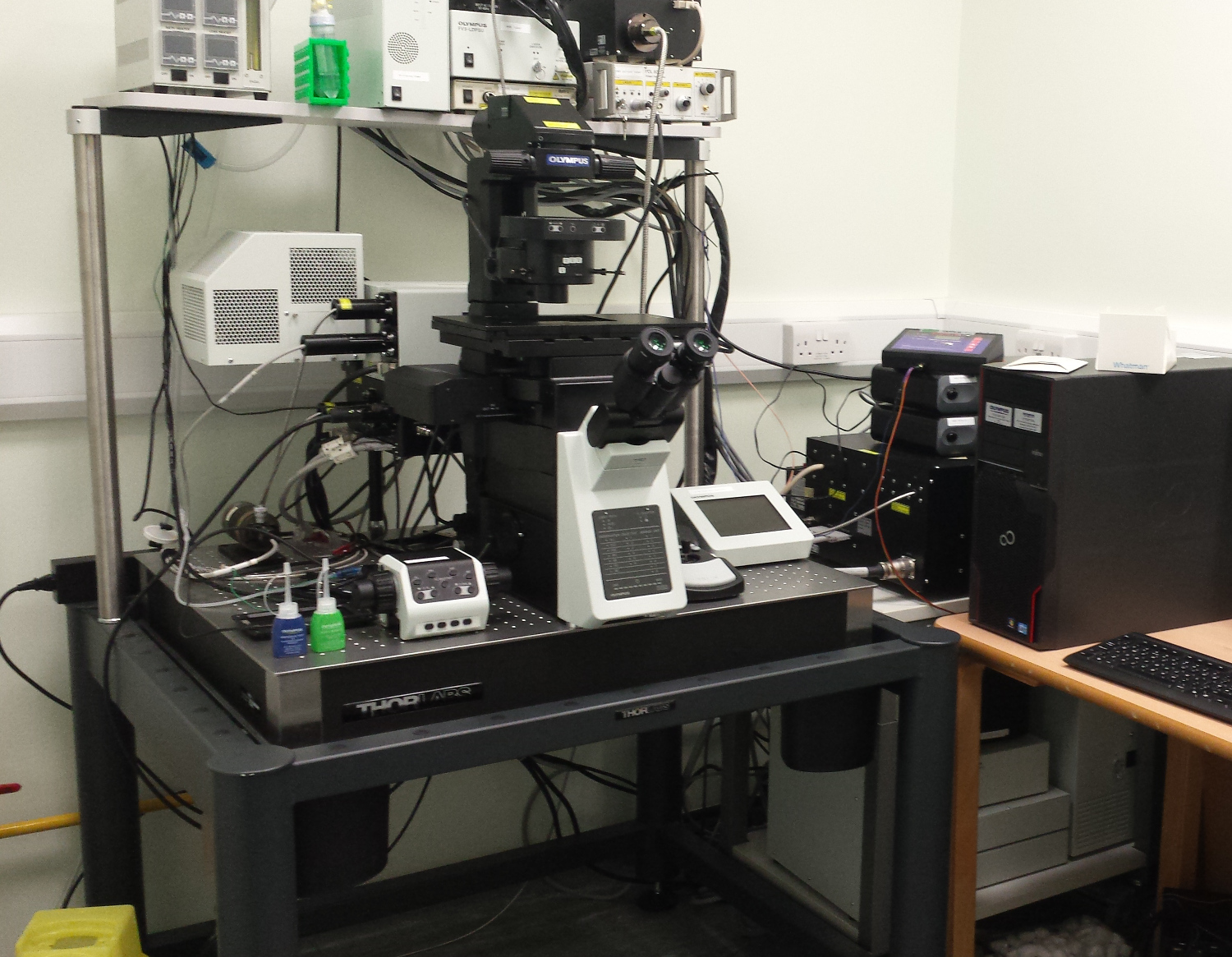
The Olympus FluoView FV1200 Confocal Laser Scanning Microscope at the LMCB allows high-quality imaging of fixed and live cells. The system is built around an IX83 inverted microscope with a fully motorized stage. The dual-fibre multi-combiner delivers 405, 440, 473, 559 and 635 nm light from diode lasers and allows the use of light for simultaneous imaging and photostimulation together with the laser light stimulation (SIM) scanner. A multi-line argon laser (458, 488 and 515 nm) is also available. There are 5 channels for fluorescence detection available, 3 standard PMTs and 2 high-sensitivity gas gallium phosphide (GaAsP) detectors for the imaging of very dim samples. The FV1200 can be configured for FRET, FRAP, and FLIP imaging, as well as laser ablation and photo-uncaging experiments. The 30x and 60x silicone oil objectives are optimized for high-resolution imaging with improved brightness.
The main galvanometer scanning mirrors are coated with an anti-oxidative layer to improve reflection efficiency for excitation and emission filters. The Z-Drift Compensation System (ZDC) allows you to carry out long time-lapse imaging without focus drift and high reliability. TokaiHit CO2 (5%) stage top incubation equipment for live cell imaging at 37 °C including a lens heating device is also available. The microscope is located in room G-09.
For further information or arranging a training session please contact: lmcb-lm-help@ucl.ac.uk